
ISSN (0970-2083)
ISSN (0970-2083)
Vadim M. Aleksandrov1*, Andrey A. Ponomarev2, Gizatulla Imashev3, Valentina E. Makhatova3, Aigul N. Shakibayeva3
1JSC "TANDEM", 625000, 57 Republic Street, Tyumen, Russian Federation
2Geochemical Laboratories in Tyumen Industrial University, 625000, 38 Volodarsky Street, Tyumen, Russian Federation
3Atyrau State University named after Kh. Dosmukhamedov, 060011, 212 Student Ave., Atyrau, Republic of Kazakhstan
Received 29 May, 2017; Accepted 04 June, 2017
Visit for more related articles at Journal of Industrial Pollution Control
At the moment, the actual direction in the chain of petrophysical drill sample analysis is an approach called "Digital core-sample" or "Digital petrophysics". However, the majority of Russian oil and gas industry companies prefer traditional methods of laboratory analysis, due to the lack of information about the possibilities of this approach. Comparative characteristics of the established laboratory methods effectiveness for determining terrigenous container rocks’ capacitive properties (Nuclear Magnetic Resonance, Preobrazhensky method; Gas Volumetric method) with the method of computer microtomography (micro-CT). Getting recommendations in the selection of test methods of open porosity evaluation in non-standard collectors – with low filtration properties to 1 mD and containing high-viscosity oils and bitumen. The open porosity (FVF) in helium was determined by the "Coretestsystems" company permeameter-porosimeter AR-608; FVF was determined by a Nuclear Magnetic Resonance (NMR) – Spectrometer Chromatec 20M, manufactured by ZAO SKB "Chromatec"; by Preobrazhensky method – according to the procedure, in the laboratory. FVF was determined by micro-CT method of "Bruker microCT" company’s microtomograph – SkyScan 1172. Comparative analysis of methods for determining container rocks’ capacitive properties has shown that standard laboratory techniques: Preobrazhensky method, Gas Volumetric Method are affordable, effective and simple. However, in the case of determining of open porosity in low-permeability container rocks containing highviscosity oil qualitative sample preparation cannot be conducted. As a result, the FVF values are underestimated. The most effective methods in the unconventional container rocks’ analysis are Nuclear Magnetic Resonance and computer microtomography. In case of a clay material high content in the container rocks, the nuclear magnetic resonance method does not give reliable results. This is confirmed by modern Russian scientists and present experimental work.
Bazhenov formation, Computer microtomography, Open porosity definition, Nuclear Magnetic Resonance method, Preobrazhensky method, Gas Volumetric method, SkySkan 1172
Information about the internal structure of the container rocks void space has been extracted in a very difficult way until recently: the specifically heated liquid agent, that solidifies after a while at room temperature, was pumped into the formation. Then the ettle was dissolved by acids: carbonate component by hydrochloric acid, silicate – by hydrofluoric acid (Khanin, 1969).
At the end of XX century the computer tomography (Shu-Yen Wan, 2002; Natterer, 2001; Mees, 2003) was introduced to the petrophysical core-samples’ researches industry, which allows to obtain information on the composition of rocks and on the structure of the void space with detalization about 150-200 microns. However, given that the pore size of terrigenous rocks begins with a hundredths of micron, a need to develop a tomographic analysis capabilities has appeared. Nowadays, nano and micro tomographs have appeared thanks to advances in technology. They allow to assess pore sizes in different scales. A new direction "Digital petrophysics" was born as a result of this.
"Digital petrophysics" or "Digital core-sample" – are a reception of petrophysical rocks’ properties by means of multi-level tomography (Vahrusheva, 2015; Alyafei, 2016; Saenger, 2016).
In this paper, we will analyze the possibilities of computer microtomography on the example of the SkyScan 1172 device – Belgium production.
Computer microtomography and micro-CT is a reconstruction of binary shadow projections of the sample into three-dimensional space.
The principle of the device’s operation is presented in (Fig. 1). Sample is installed into microtomograph on the object table, which rotates during scanning. At this point, X-rays emitted by the source pass through the sample, some of which are absorbed by the object of study. The receiver (camera) captures shadow projections of the sample. A separate projection is being fixed at every turn.

Fig. 1 Computer tomohraphy method, principles of work.
Brightness (different shades of gray) on the X-ray shadow projection reflects the weakening of the X-rays due to scattering and absorption effects of the signal transmitted through the sample.
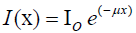
The attenuation will depend on the thickness and density of the studied object, as well as on the effective atomic number (Zeff), which the test sample consists of. This effect is described by the Beer-Lambert law which determines the attenuation of monochromatic light beam propagating in an absorbing medium. The law is represented by formula:
 (1)
(1)
where l0 is the initial X-ray intensity; x – thickness of the material’s layer through which the radiation passes; μ– indicator of the medium’s absorption.
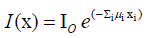
In case when the scanned object is not homogeneous in composition, such as almost all rocks, the equation becomes:
 (2)
(2)
where a separate material with a specific absorption index of μi and linear length of xi corresponds to each increment of i. In the geological objects study when using radiation sources with energy up to 100 keV, the main process determining the attenuation of X-rays is the photoelectric absorption (Stuart, 2009).
After scanning in the course of reconstruction, the projections are converted into thick one pixel slices. The sample’s 3D model is constructed and analyzed from them. Maximum sample size for 1172 SkyScan system should not exceed 64 mm length/widthwise. Maximum height – 70 mm (Ponomarev, 2015).
Existing and methodically tuned laboratory infrastructure of petrophysical core-samples research dictates its own laws, which are difficult to refuse. Methods of porosity determination in conventional reservoirs have been developed and tested for decades, and are not to be questioned. Currently, however, due to the depletion of oil reserves, there is a need to develop a deposit with hard recoverable reserves.
For clarity, lithotypes of Bazhenov Formation, which are the collectors, were chosen as research objects (based on AO of Siberian Research Institute of Petroleum Industry):
a) clay-siliceous rock until silicite (silicon content more than 50%), biomorphic, rich in organic matter (OM); Apatite content is characterized up to 50% and carbonate up to 20%.
b) sandwiching of calcareous phosphate oil- saturated rock (high value of P2O5 more than 17%) and mudstone rich in OM. Characterized by the clay content up to 35% (Fig. 2).

Fig. 2 Lithotypes of Bazhenov formation, which are the collectors: ÃÂ – Tonquarz until silicit; B – Sandwiching of calcareous phosphate rock.
The experiment consisted in obtaining comparative characteristics of determining the porosity of nonstandard container rocks by methods of: Nuclear Magnetic Resonance (NMR); Preobrazhensky method; Gas Volumetric method and computer microtomography (micro-CT).
NMR
Using the effect of Nuclear Magnetic Resonance (NMR) to determine the characteristics of the subsurface rocks based on the possibility of a direct indication of water content of pore fluids (water, oil, gas) which are directly in the pore space. When NMR studying of the pore space of subsurface rocks the reaction of pore fluid magnetization on resonance effect is measured, that causes the precession of the hydrogen nuclei in a magnetic field.
Preobrazhensky method
Porosity is determined by saturating the rock with kerosene or 3% solution of salt water. It is determined by the difference in the weight of dry and saturated sample, ascribed to the volume of the sample, multiplied by the density of the saturating fluid. The ratio of pores’ volume to the sample’s volume gives the required value of the porosity. It is expressed in % or as a unit fraction.
Gas Volumetric method
To determine the coefficient of open porosity by using Gas Volumetric method such formula was used
CoP = (Vev – Vsc) / Vev (3)
where Vev – external sample’s volume, Vsk – volume of the sample’s mineral skeleton.
Determining the external volume was conducted on the basis of sample’s geometrical sizes measurement using calipers with digital readout. To reduce the error in determining Vev, the measurement of geometrical sizes was conducted in three locations and averaged.
Micro-CT method
Porosity was determined by "Skyscan 1172" microtomograph with such scan settings: Filter: Al+Cu; angle of rotation – 0.3; the number of frames – 4; the number of random frames – 5; scanning at 360 degrees; resolution of 2.9 microns per pixel; software – "NRecon", "CTan". Isolation of pores by X-ray density intervals of the interpreter is conducted similarly to the analysis of lithological slides under the microscope, but micro-CT is characterized by high detail.
The For the results of the open porosity determining of Bazhenov’s Formation collectors (Table 1).
| Sample | Gas Volumetric | NMR | Preobrazhensky | Micro-CT |
|---|---|---|---|---|
| ÃÂ | 11.79 | 13.7 | 4.2 | 13.8 |
| B | 3.46 | 8.39 | 0.7 | 16.2 |
Table 1. The results of defining FVF by methods of NMR; Preobrazhensky; Gas Volumetric; Micro-CT
As you can see, the results of the open porosity defining have turned out quite different. In order to understand the reasons for this, it should be noted that the permeability of these samples does not exceed 1mD. Furthermore, geochemical analysis of organic substance indicates high content of bitumoid and high viscosity oil in samples. In this regard, it is worth paying special attention to the fact that before Gas Volumetric analysis and the determination of open porosity by Preobrazhensky method – samples undergo special sample preparation – extraction of oil from the sample is conducted with an organic solvent. According to the results it can be stated that it is impossible to completely remove the oil from the rock without destroying it while defining the open porosity of low permeability container rocks containing high-viscosity oil. The most effective methods are: NMR and micro-CT – open porosity values per "A" sample confirm this.
However, in the "B" sample there is a significant discrepancy between the results of micro-CT and NMR methods. Analysis of the literature on the operating principle of the method of Nuclear Magnetic Resonance indicates the low efficiency in the determination of the petrophysical properties of rocks with a high content of clay (Shumskayte, 2015; Zubkov, 2014).
The Comparative analysis of methods for the determination of reservoir properties of container rocks has shown that the standard laboratory techniques: Preobrazhensky method, Gas Volumetric method – are very affordable, effective and simple. However, in case of open porosity defining in low-permeability container rocks containing highviscosity oil it is impossible to produce high-quality sample preparation. As a result, the values of CoP obtained are underestimated.
The most effective thing in this case is to apply Nuclear Magnetic Resonance and computer microtomography techniques.
In case of a high content of clay material in container rocks, the Nuclear Magnetic Resonance method does not provide reliable results. This is confirmed by modern Russian scientists and present experimental work.
Copyright © 2024 Research and Reviews, All Rights Reserved