
ISSN (0970-2083)
ISSN (0970-2083)
Uttam Kumar1 and V.K. Mishra2*
1Department of Biotechnology, Doon P.G. College of Agriculture Science and Technology, Dehradun 248 001, Uttaranchal, India
2Department of Biotechnology, Doon P.G. Paramedical College,Dehradun 248 001, Uttaranchal, India
Received date: 15 April, 2015; Accepted date: 17 June, 2015
Visit for more related articles at Journal of Industrial Pollution Control
Orange G, asulfonated azo dye, is used in silk, wool products and dyeing. It is recalcitrant to oxidative degradation, and persists in aquatic environment. In the present study, Orange G dye degrading bacterial strain was isolated from soil containing rusted iron shavings. The present study illustrates the ability of a bacterial strain S2, to degrade the Orange G. The strain was identified as Bacillus sp. based on chemical and phylogenetic analysis. The study presents comparative assessment of degradation of Orange G by free and immobilized cells of Bacillus sp. S2. The dye degradation was confirmed through UV-visible spectroscopy, HPLC and GC-MS analysis. Spectral analysis of the product following dye degradation showed that the medium did not contain any aromatic compounds. In the decolorization study, the strain showed 100% degradation of dye at 100 mgL-1 dye concentration by free cells at 24 h incubation, pH 7.0 and temperature 37o C whereas immobilized Ca-alginate beads with strain showed 100 % degradation up to 250 mg L-1 of dye concentration under similar conditions. In packed bed reactor, highest dye degradation was obtained at lower flow rate. The study could be used for the development Ca-alginate immobilized bacterial cells in wastewater treatment plants.
Ca-alginate, Degradation, GC MS, HPLC, Immobilization, Orange G, PCR, 16S rDNA
Synthetic organic colorants are used universally in manufacturing processes ranging from food and textile production to the printing and pharmaceutical industries (Carmen and Daniela, 2010). Some of these dyes, their precursors, or their biotransformation products such as aromatic amines, have been shown to be mutagenic and carcinogenic (Chung, 1983). There are more than 10,000 of different dyes and pigments used industrially, and over 0.7 million tons of synthetic dyes are produced annually worldwide (Robinson et al., 2001). Among all the available synthetic dyes, azo dyes are the largest group of dyes used in textile industry constituting 60-70% of all dyestuffs (Saratale et al., 2011). About 10-25% of textile dyes are lost during dyeing process, and 2-25% are directly discharged as water effluents in different environmental components (Carmen and Daniela, 2010). Unfortunately, most of the dyes escape conven tional wastewater treatment processes and persist in the environment as a result of their high stability against light, temperature, water, detergents, chemicals, and microbial attack (Couto, 2009). In particular, the discharge of dye-containing effluents into the water environment is undesirable, not only because of their colour, but also because many of dyes released and their breakdown products are toxic, carcinogenic or mutagenic to life form (Zaharia et al., 2009). Currently, implementation of physico-chemical methods, such as adsorption, electrocoagulation, flocculation, froth floatation, ion exchange, membrane filtration, ozonization, and reverse osmosis, have met with limitations of being high operational cost, inability to completely remove the recalcitrant azo dyes and/or their organic metabolites and the secondary pollutants, and huge amount of sludge generation. Therefore, dye degradation using microorganisms is an attractive strategy in biodegradation since it is an eco-friendly and cost-competitive alternative to chemical decomposition process (Saratale et al., 2010).
The biodegradation of azo dyes may occur either aerobically, anaerobic or by a combination of both (Solís et al., 2012). Reductive cleavage of azo bonds (–N=N–) by azoreductase results in formation of aromatic amines. Some of these aromatic amines are known to be cytotoxic, mutagenic and carcinogenic. These toxic substances are removed by aerobic treatments. Thus, sequential anaerobic/aerobic systems can be effective in complete mineralization of dye (Popli and Patel, 2015). If the aerobic and anaerobic systems designed in a single unit, such as immobilized cells in a tubular bed reactor, dye removal systems would be simplified.
Orange G, asulfonated azo dye, is used in silk, wool products and dyeing, also can dye paper and manufacturing ink, used in wood and biological dyeing. There are only few reports of degradation of Orange G (Tripathi and Srivastava, 2011). So far there are limited reports of degradation of Orange G by immobilized system are available. This study presents batch degradation of Orange G by immobilized Bacillus sp. S2.
Chemicals
The textile dye Orange G (C.I.16230) was obtained from Himedia, India and Fig. 1 shows the structure of dye.

Figure 1: Chemical structure of Orange G
Sample collection
Rusted iron scraps was collected from waste-dump site from Selakuai (geographic site location: Longitude 77.84 E, Latitude 30.34 N, Uttarakhand, (India) and was used for isolation of Orange-G azo dye degrading bacteria.
Medium
Nutrient broth and agar, Mineral Salt media (MSM) with composition (per litre distilled water) : K2HPO4 -1.0 g , KH2 PO4 -1.0 g, MgSO4 .7 H2O- 0.2g, CaCl2- 0.2g, FeCl3.6H20- 0.05 g, NH4 NO3 - 1.0 g.
Isolation and characterization of dye degrading bacteria
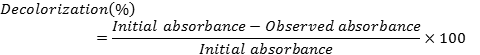
Small portions of rusted iron scraps pieces (about 1 g) were inoculated into 100 mL pre-sterilized Mineral salt medium (MSM) (pH 7.0) with yeast extract in a 250 mL flask containing Orange G (100 mgL-1). Then flasks were incubated at 37oC on a rotatory shaker at 120 rpm. Pure culture of each isolate was maintained on MSM with 1% agar. A loopful of culture was inoculated on MSM medium with 100 mgL-1 of dye, and was incubated at 37oC on a rotatory shaker at 120 rpm. After every 6 h of interval, screening of dye decolorizing bacteria was carried out by monitoring absorbance at 480 nm. The isolate that was showing fastest decolorization of dye, was selected and designated as S2 strain. Decolorization activity was calculated as follows:
Morphological characterization and biochemical tests for preliminary identification of Orange G dye degrading bacteria were performed for according to Bergey's manual of systematic bacteriology (Williams et al., 1993). For identification by 16S rRNA gene sequence analysis, genomic DNA was isolated using a fast DNA kit (Q-Biogene), and was amplified by polymerase chain reaction (PCR) using primer sets Primers: pA (5'-AGAGTTTGATCCTGGCTCAG-3') and pH (5'-AAGGAGGTGATCCAGCCGCA-3'). 50 microlitre reaction mixture 100 ng of total DNA, 2 U of Taq polymerase, 0.2mM of dNTPs 3.0 mM of MgCl2 and 0.4 μM of each primer. The PCR amplifications were carried out using initial denaturation step of 10 min at 94oC, followed by 30 cycles 1 min 94oC, 30s 55oC, 72oC for 1 min. The reaction was completed at final extension temperature 72oC for 10 min. The presence of PCR products was determined by electrophoresis of 10 μL of the reaction product in a 0.8 % agarose gel.
The PCR product was sequenced at Ocimum Biosolution (P) Ltd. (India). Identification was achieved by comparing the 16S rRNA genes sequences obtained with other 16S rRNA genes in the GenBank using the BLASTn program. The phylogenetic tree was constructed by using the neighbor-joining (NJ) analysis method. Further, the Bootstrap analysis was performed to assess the confidence limits of the branching. The sequence was deposited in Gene Bank with accession no. JX125394.
Immobilized cell preparation
3% cell pellet of Bacillus sp. 2 was entrapped in 3% Ca-alginate solution using ice cold, 0.2 M CaCl2 solution. After 30 min of bead formation, it was washed with 0.85 % NaCl and stored at 4oC until further use.
Batch Degradation experiments using free and immobilized cells
For degradation with free cells, 5mL of inoculum (8 ×10⁸ CFU/mL) of isolate S2 was added to a 250 mL of flask containing 100 mL of MSM medium (pH 7.0) with 15 mg yeast extract, and different concentrations of Orange G (100, 150, 200, 250, 300, 350, 400, 450 and 500 mgL-1). The flasks were placed in a rotary shaker, agitated at 180 rpm at 37°C for 48 h.
For degradation with immobilized cells, 5 g (fresh weight) of Ca-alginate beads cells were placed in 250 mL of conical flask containing 100 mL of MSM medium with yeast extract, and Orange G was added at different concentration, (100, 150, 200, 250, 300, 350, 400, 450 and 500 mgL-1). The flasks were placed in a rotary shaker at 180 rpm at 37°C for 48 h.
Continuous degradation experiment in packed bed reactor
For continuous degradation, dye decolorization was carried out in a packed bed reactor with immobilized Ca-alginate beads 2% (w/v). The continuous supply of sterile MSM medium, with yeast extract (pH 7.0) and Orange G, was made with the help of a peristaltic pump. The reaction temperature was controlled at 37 °C using a water bath.
HPLC analysis
After decolorization, the medium was analyzed through HPLC 1260 infinity Agilent using Eclipse Pluse- C18 column (4.6 mm x 100 mm x 5.0 μm). The mobile phase was composed of 40% water (v/v) and 60 % acetonitrile (v/v). 10 μL of sample was injected. The compounds were eluted at a flow rate of 1.0 mL/ min. UV-Vis detector was used for the determination of Orange G dye content in control and treated samples. The photodiode array detector was set at 483 nm for colour detection due to dye in visible range, and to detect changes in UV absorbance at 248 nm. The elution was monitored at 248 nm, and coloured products at 483 nm. The percentage of degradation was calculated by comparing the peak area of degraded and control sample.
GC-MS analysis
Following decolorization, the sample was extracted with dichloromethane and dried over anhydrous Na2SO4 and evaporated to dryness. The extracted sample was analyzed through GC-MS Shimadzu Mass Spectrometer-2010 series. The sample was injected in GC-MS equipped with a split injector and a PE Auto system XL gas chromatograph interfaced with a Turbo-mass spectrometric mass selective detector system. The MS was operated in the EI mode (70 eV). Helium was employed as the carrier gas and its flow rate was adjusted to 1.2 mL/min. The analytical column connected to the system was an Omega Wax capillary column (length- 30m × 0.25 mm i.d., 0.25 μm film thickness). The column head pressure was adjusted to 90.4 kPa. Column temperature programmed from 80 oC (2 min) to 200 oC at 10 oC/min and from 200o C to 235 oC at 15 oC/min with hold time 5 and 10 min respectively. A solvent delay of 4 min was selected. The injector temperature was set at 270 °C. The GC-MS interface was maintained at 280 °C. The MS was operated in the acquisition mode scanning from m/z 40 to 650.0. In the full scan mode, electron ionization (EI) mass spectra in the range of 40- 650 (m/z) were recorded at electron energy of 70 eV. The metabolic intermediates from Orange-G degradation were identified by comparing their retention time (RT in min) and mass spectra with library of the National Institute of Standard and Technology (NIST), USA/Wiley.
Isolation and characterization
The bacterial strain, S2 showing remarkably fast Orange- G decolorization capacity, was isolated from rusted iron materials (Selakuai, Dehradun, India). It is reported that rusted iron materials are rich in bacteria (Khan et al., 2010) The strain S2 was found superior to other isolates on several criteria, i.e. fast decolorization rate, ability to tolerate and degrade high concentration, and capacity to degrade range of dyes. Morphological and biochemical characterization of the strain S2 is presented Table 1. The strain S2 was a rod shaped gram negative bacteria capable of utilizing broad range of carbohydrate, i.e. sucrose, glucose and fructose; but could not utilize xylose, cellobiose, mannitol and galactose. 16S rRNA gene sequence analysis is most widely used method for the molecular identification and differentiation of bacterial species. 16S rRNA gene sequence of the strain was amplified by polymerase chain reaction (PCR). In phylogenetic analysis, the strain S2 has closest relatedness with that of Bacillus sp. 21059 (GQ199761) (Fig. 2). Based on sequence similarity and blast analysis, bacterial strain S2 was identified as Bacillus sp., and partial 16S rRNA gene sequence of the strain was deposited in GeneBank with accession number JX125394, Bacillus sp. S2.

Table 1: Morphological and biochemical characterization of strain S2 from rusted iron scraps

Figure 2: Phylogeny tree of the strain, Bacillus sp. S 2 for the partial sequence based on 16S rDNA
Dye Degradation
Orange G dye degradation by free bacterial cells have been reported by few investigators (Shah, 2014). In this study, we present decolorization of Orange G by free and immobilized cells of Bacillus sp. S2. (Fig. 3). In the decolorization study at 24 h, our strain showed 100% degradation of dye at 100 mgL-1 dye concentration by free cells whereas immobilized Ca-alginate beads with strain showed 100 % degradation up to 250 mg L-1 of dye concentration (Fig. 3). At 250 mg L-1 of dye, free cells only showed 45% of dye decolorization of incubation while immobilized cells showed 100% dye decolorization during the same time of incubation (Fig. 4).

Figure 3: Degradation of Orange G by free and immobilized Ca-alginate beads of Bacillus sp. S2 in batch culture

Figure 4: Time course decolorization of Orange G by free and immobilized cells at 250 mgL-1 dye concentrations
During repeated use of Ca-alginate beads, there was leakage of cells from gel matrix. After 10 cycles, cell leakage to the extent 1 × 10⁵ was observed. Compared to free cells, immobilized system is more stable for industrial processes and allows it to be reused many times. There are reports of use of Ca-alginate immobilized bacterial cells to reduce azo bonds enzymatically and used as a biocatalyst for decolourization of azo dye (Usha et al., 2012), and azo dye precursor (Usha et al., 2010). However, there is no report available on degradation of Orange G by immobilized Ca-alginate bacterial cells. The Ca-alginate tubular gel provides both anoxic and aerobic system: within the gel- anoxic center; and the outer surface of the gelaerobic centre, for degradation of aromatic amines formed from reductive cleavage of azo bond (Ogugbue et al., 2012). In packed bed reactor, dye decolorization decreased significantly with increasing dye concentration. Efficiency of dye decolorization was better at lower flow rate (Fig. 5). In our study, we report a simple and efficient- one step system for decolorization of Orange G in a Ca-alginate immobilized packed bed reactor.

Figure 5: Effect of Orange G concentrations and flow rates on decolorization of dye by Bacillus sp. S2 in packed bed reactor reactor
The azo dye degrading ability of the strain was further confirmed by UV-Vis spectrophotometry, HPLC, and GC-MS analysis. UV-Vis analysis revealed decrease in the intensity of major peak (λmax 480 nm) in visible region, and shift in the peaks in UV region (Fig. 6). Spectral changes of the input and output of the bioreactor by UV-visible spectroscopy indicated decolorization of the dye solution. The first step in the bacterial degradation of azo dyes is the reduction of the –N=N– bond by azoreductase which results in decolorization (Ogugbue et al., 2012).

Figure 6: UV-Vis spectral analysis of Orange G decolorization by Bacillus sp. S2
The dye decolorization and biodegradation was fur-ther confirmed by HPLC analysis. In visible region, intensity of color was monitored at 483 nm. In treated sample (bacteria with dye), the peak at 483 nm disappeared completely. However, new peaks at retention time RT 2.407, 2.553 and 2.740 appeared in treated sample that were not present in the control (broth with Orange G dye without bacteria) (Fig. 7). In UV region, a single peak of Orange G dye was present in control with retention time 2.067 when peak intensity monitored at 248 nm. However, treated test sample (dye with bacteria) showed formation of new three peaks with RT 2.153, 2.287 and 2.640 indicating degradation of dye (Table 2). GC-MS analysis of degraded sample by Bacillus sp. S2 in extractable products showed that in control sample, only Orange G (RT=8.909) is identified (Fig. 8a), whereas in degraded sample different intermediates were identified using the NIST mass spectral database: formation of phenyl hydrazine (RT=12.654), aniline (RT=13.535) (Fig. 8b). However, some other peaks in the chromatogram remained unidentified. Difference in peaks of the input and output of the bioreactor indicated decolorization of the dye solution by degradation. Spectral analysis of the products after Orange G utilization showed that the spent medium did not contain any aromatic compounds. The aromatic amines can be mineralized by means of aerobic treatment by non-specific enzymes via hydroxylation and ring-opening in the presence of oxygen (Pandey et al., 2007). The reductive cleavage of the azo bond dissipates the electron deficiency of the aromatic nuclei so that the aromatic amino compounds generated may be amenable to subsequent oxidation and mineralization (Forgacs et al., 2004). The results obtained in the present investigation is corroborated with the finding of Shah (2014) that indicated formation phenyl hydrazine which further converted to aniline followed by aromatic ring cleavage leading to complete mineralization of Acid Orange 10. Both anoxic and aerobic conditions are required for degradation of azo dye (Mahmood et al., 2015).

Table 2: HPLC analysis with detector at 248 nm in UV region
There are very few reports on the biodegradation and detoxification of textile and dye-stuff industrial wastes containing Orange G. In this study, we describe the isolation and characterization of of Bacillus sp. S2 capable of efficiently degrading Orange G dyes in free and immobilized system.
Copyright © 2026 Research and Reviews, All Rights Reserved